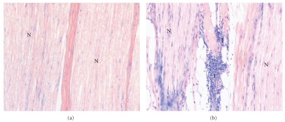
Figure 3.
(a) Longitudinal microscopic view (×200, Giemsa stained) of the radial nerve after needle placement by means of nerve stimulation. A minimal threshold current of 1.0 mA was applied for needle positioning. The needle did not contact the nerve tissue. N, nerve fascicle; I, inflammatory cells. Score value, 0 (b) Longitudinal microscopic view (×200, Giemsa stained) of the median nerve after needle placement by means of nerve stimulation. A minimal threshold current of 0.2 mA was applied for needle positioning. The needle contacted the nerve tissue. N, nerve fascicle; I, inflammatory cells. Score value, 2.0.

