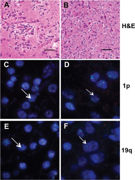
Figure 1. H&E histology and FISH of 1p19q nondeleted or codeleted WHO grade II oligodendrogliomas.
Representative H&E photomicrographs and FISH analysis for 1p/19q nondeleted (A, C, E) and 1p/19q codeleted (B, D, F) are shown. The H&E sections (A, B) demonstrate typical morphologic features of WHO grade II oligodendrogliomas for both genotypes which are indistinguishable on histologic grounds. However, FISH analysis discriminates between these tumors based on the ratio of centromeric (green probes) and subtelomeric 1p (C, D) and 19q (E, F) specific probes (red signal), which is 2:2 in the nondeleted tumor (C, E) and 2:1 in the codeleted tumor (D, F). Representative cell nucleus is shown with long and short arrows, respectively. Scale bar 50 μM.

