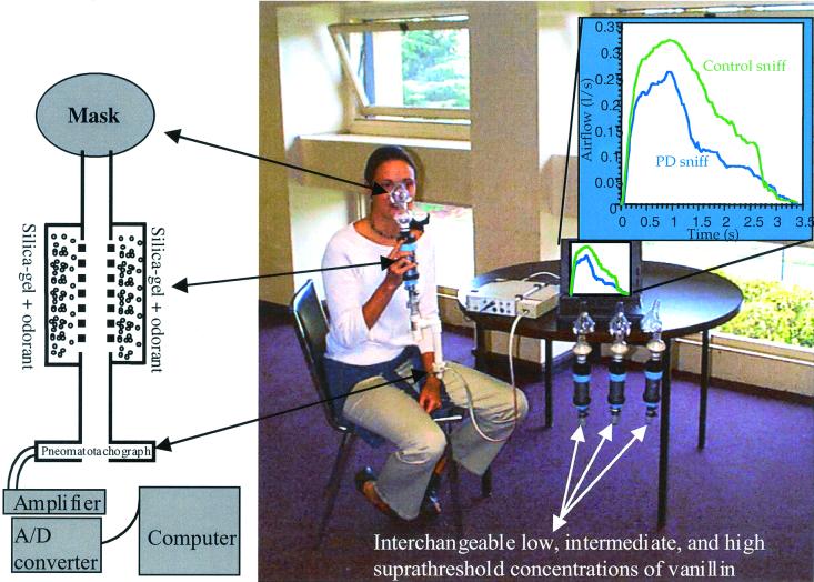
Figure 1.
Methods for recording airflow. A schematic drawing and image of the recording apparatus as used in refs. 22 and 27 (note the three additional interchangeable odorant sources at the table). Subjects were told that the tubes connected to the mask were the odorant supply (they were in fact the pressure transduction tubes). Subsequent questioning revealed that no subject was aware of the ongoing airflow recording. The data in the upper right quadrant is the actual mean first sniff of 20 patients and 20 controls (the person demonstrating the apparatus is not a patient).

