
Figure 1.
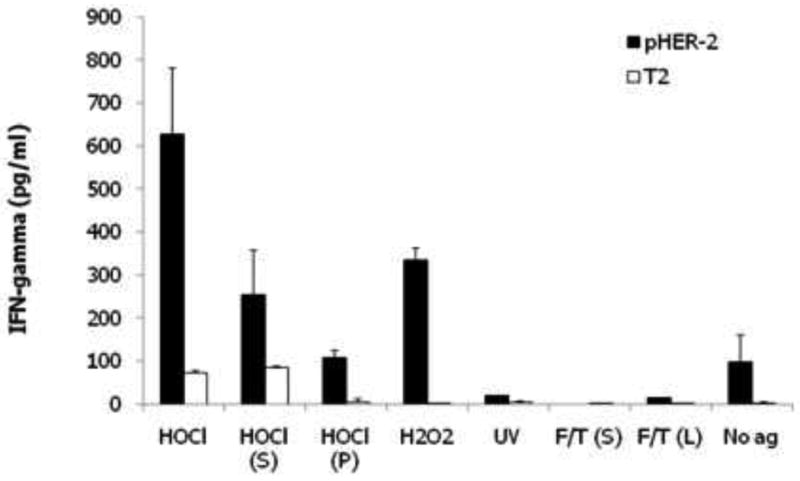
Comparison of immunogenicity of human DCs pulsed with different lysate preparations of SKOV3 ovarian cancer cells. Autologous DC from HLA-A2+ donor, derived from elutriated peripheral blood monocytes treated with GM-CSF and IL-4 for 48 hours, were pulsed with lysates of SKOV3 cells, which express Her2, for 4 hours and matured overnight with LPS and IFN-γ. DCs were then used to prime naive, autologous lymphocytes for 10 days. Output T cells were incubated overnight with T2 cells pulsed with HLA-A2 restricted Her2 peptide or unpulsed T2 cells. The relative presence of functional Her-2 reactive lymphocytes was measured by IFN-γ ELISA. DCs were pulsed with freeze-thaw lysates of HOCl-treated whole tumor cells (HOCl); supernatants [HOCl (S)] or pellets [HOCl (P)] of freeze-thaw lysate of HOCl-treated tumor cells; freeze-thaw lysates of H2O2-treated whole tumor cells (H2O2); supernatants of freeze-thaw lysate of UVB-irradiated tumor cells (UV); and supernatants [F/T (S)] or whole freeze-thaw tumor cell lysate [F/T (L)]. Unpulsed DCs (No ag) were also matured with LPS and IFN-γ. Error bars represent data from duplicate co-culture wells. T cell priming against Her2 was highest in T cells incubated with DCs pulsed with HOCl-oxidized whole tumor cells.