
Figure 2.
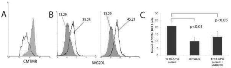
Activation of mouse DCs by tumor cells killed with oncolytic replication-restricted HSV. (A) Tumor-infiltrating CD8+ T cells isolated from ID8 tumors were incubated with bone marrow derived DCs that were pulsed with ID8 ovarian tumor cells killed by HSV-1716 (clear) or DCs pulsed with UV-irradiated ID8 cells (gray). DCs pulsed with viral oncolysate induce more pronounced proliferation of CD8+ T cells, as assessed by CMTMR dilution. (B) Flow cytometry analysis of NKG2D ligand (NKG2DL) expression in (left) unpulsed immature DCs (gray) or unpulsed DCs matured with LPS (clear), and (right) control unpulsed DCs (gray) and DCs pulsed with ID8 cells killed by HSV-1716 (clear). NKG2DL was detected through NKG2D/FC chimera (R&D). (C) Percent of CD25+ activated NK cells as determined by flow cytometry analysis 48 hours after incubation with immature DCs or DCs pulsed with apoptotic cells killed with HSV-1716 (1716-APO), in the presence or absence of soluble NKG2D (5 g/ml).