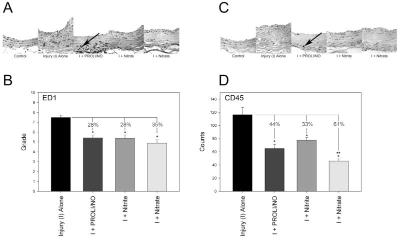
Figure 4.
Inflammation following rat carotid artery injury at 14 days: Representative sections and quantification of (A and B) monocyte/macrophage (ED1) and (C and D) leukocyte (CD45) infiltration, respectively. ED1 staining was quantified on a scale of 0–4 for the intima, media, and adventitia, and the sum of these grades is reported. CD45 staining was quantified as the number of positive staining cells (e.g., at arrow) per high power field. *p<0.05 vs. injury alone. **p=0.035 vs. nitrite. I=injury.

