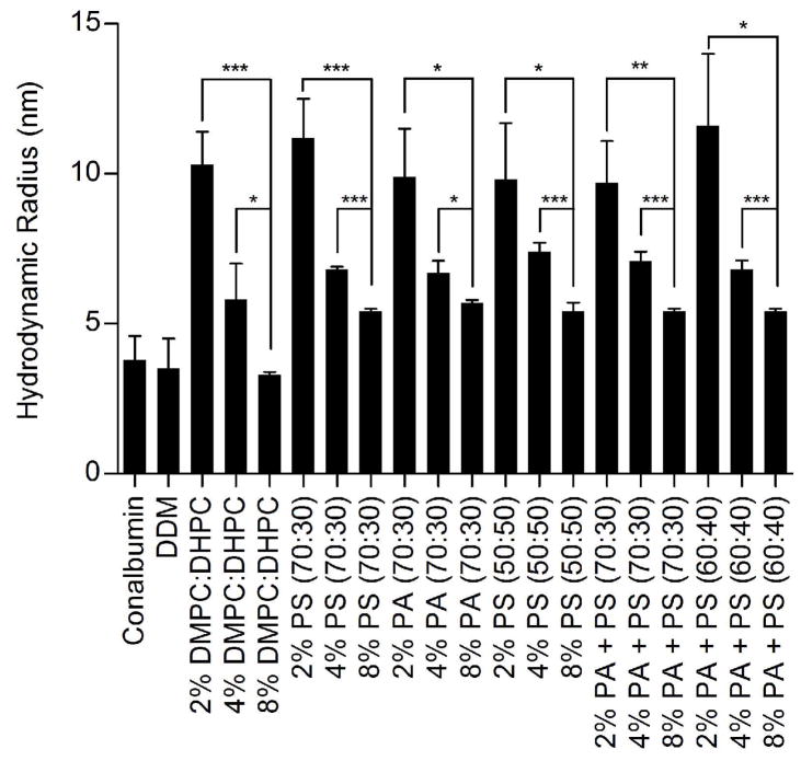
Figure 5.
Dynamic Light Scattering measurements on negatively charged bicelles. Data was collected on samples at final phospholipid concentrations of 2%, 4%, and 8% in solution. All samples were prepared in extra MII assay buffer (50 mM HEPES pH 8.0, 100 mM NaCl, 1 mM MgCl2). 2 mg/mL Conalbumin (75 kDa) and 0.5 mM DDM (70 kDa) were prepared as positive controls. Hydrodynamic radii were determined by Dynamics V5 software, with light scattering data collected at 18–22°C on a DynaPro detector. The ratio of neutral to negatively charged phospholipids is indicated within the parentheses in the graph. Results are are means ± S.E.M. of at least 25 scans with two independent experiments (* p < 0.05; ** p < 0.01; *** p < 0.001; NS, Not Significant).

