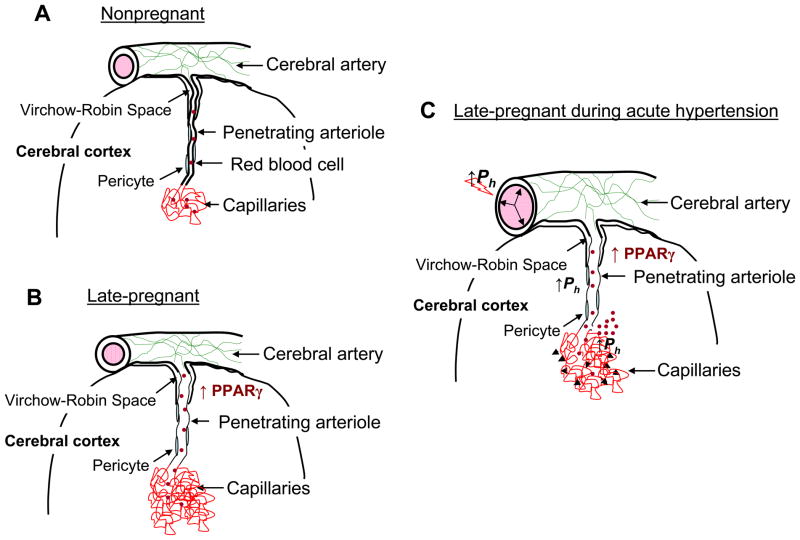
Figure 4. Summary diagram of cerebral vascular adaptation to pregnancy and the effect of acute hypertension.
(A) Cerebral arteries and arterioles that lie on top of the brain give rise to smaller arterioles that penetrate into the brain tissue, passing first through the Virchow-Robin Space. After passing through this space, arterioles are closely associated with multiple cell types, including pericytes, astrocytes and neurons. Penetrating arterioles are long and largely unbranched segments of the vasculature that connect upstream vessels to the capillary microcirculation and as such contribute significantly to cerebrovascular resistance in the brain. (B) During pregnancy, penetrating arterioles undergo outward hypotrophic remodeling under the influence of PPARγ activation that is increased during pregnancy. In addition to outward remodeling of arterioles in the brain, capillary density increases. These alterations in structure appear to occur only in the vasculature associated with brain parenchyma, a segment of the vasculature in which expression of PPARγ is relatively low. Thus, we speculate that PPARγ-dependent mechanisms in brain parenchyma have a paracrine influence on the associated vasculature. We further speculate that upstream cerebral arteries, that appear unaffected by pregnancy and PPARγ activation, maintain vascular resistance that is protective in downstream microvessels in relation to hydrostatic pressure. (C) During acute hypertension, similar to what occurs during severe preeclampsia and eclampsia, forced dilatation of large cerebral arteries occurs, decreasing vascular resistance and allowing greater transmission of hydrostatic pressure to downstream arterioles and capillaries. Because the arterioles have undergone outward hypotrophic remodeling, wall stress is significantly elevated, an effect that could promote increases in permeability as well as rupture and hemorrhage (denoted in the figure by the black arrows). Increases in hydrostatic pressure also affect the capillary bed to increase transcapillary filtration and promote edema formation that is greater during pregnancy due to decreased vascular resistance and increased vascular volume and capillary density. Published as Cipolla MJ, et al. J Appl Physiol. 2010 Am Physiol Soc., used with permission.

