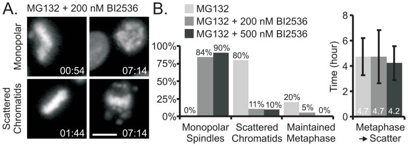
Figure 3.
Inhibition of Plk1 in metaphase-arrested cells does not block cohesion fatigue. (A) Live cell imaging indicated that HeLa H2B-GFP cells arrested in mitosis by proteasome inhibition (MG132) and treated with the Plk1 inhibitor, BI2536 at 200 or 500 nM, either formed monopolar spindles, or they maintained metaphase arrest that typically led to cohesion fatigue. (B) Quantification of HeLa H2B-GFP cells treated with MG132 and BI2536: Cells treated with only MG132 maintained bipolar spindles; 80% of these cells experienced cohesion fatigue with an average metaphase to scatter duration of 4.7 ± 1.4 hours. While the majority of cells treated with MG132 and BI2536 formed monopolar spindles, some maintained bipolar spindles, most of which experienced cohesion fatigue with an average metaphase-to-scatter duration similar to that of cells in MG132 alone. Bars = 10 μm, Time = hr:min, Error bars = standard deviation (see also Figure S3 and Movie S5)

