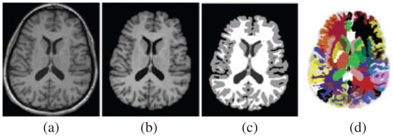
Fig. 1.

Segmentation of the brain using HAMMER in a representative T1-weighted axial image. (a) Original image. (b) Skull-removal outcome using skull strip (http://idealab.ucdavis.edu/index.php) followed by BET. (c) Outcome of segmentation into WM, GM and CSF using FSL FAST. (d) Colour-coded anatomical segmentation generated by HAMMER.
