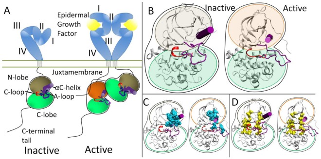
Figure 1.
(A) Activation scheme for the ErbB family. The inactive kinase (brown N-lobe) is auto-inhibited through the A-loop and αC-helix (purple). Introduction of the asymmetric dimer interface rotates the αC-helix to the active state (orange N-lobe). (B) Enhanced view of the inactive and active kinase domains. (C) Hydrophobic core (cyan) in the inactive and active conformations. (D) C-spine (left yellow spine) and R-spine (right yellow spine) in the inactive and active conformations.

