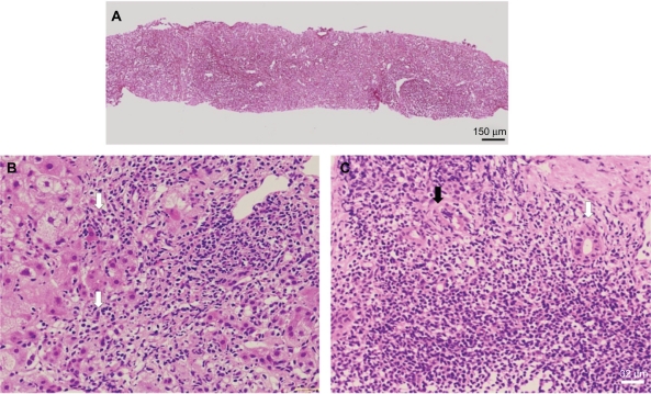
Figure 2.
Needle biopsy specimen of the liver.
Notes: A) Low magnification of silver impregnation staining for reticulin showing broad areas of parenchymal collapse. B) High magnification of a hematoxylin and eosin stained section showing interfacial hepatitis (arrow). C) High magnification of a hematoxylin and eosin stained section showing non-destructive cholangitis (black arrowhead) and destructive cholangitis (white arrowhead) surrounded by numerous plasma lymphocytic cells.

