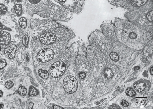
Figure 3.
Electron microscopy findings.
Notes: Migrating lymphocytes were found more frequently in a medium-sized interlobular bile duct surrounded by many inflammatory cells. One lymphocyte had just migrated through the bile duct epithelial cells. Complete absence or partial loss of microvilli and microvillous bleb formation was visible in the same bile duct epithelial cell. The arrow indicates bleb formation. The arrowhead denotes migrating lymphocytes through the bile duct epithelial cells.

