
Figure 1.
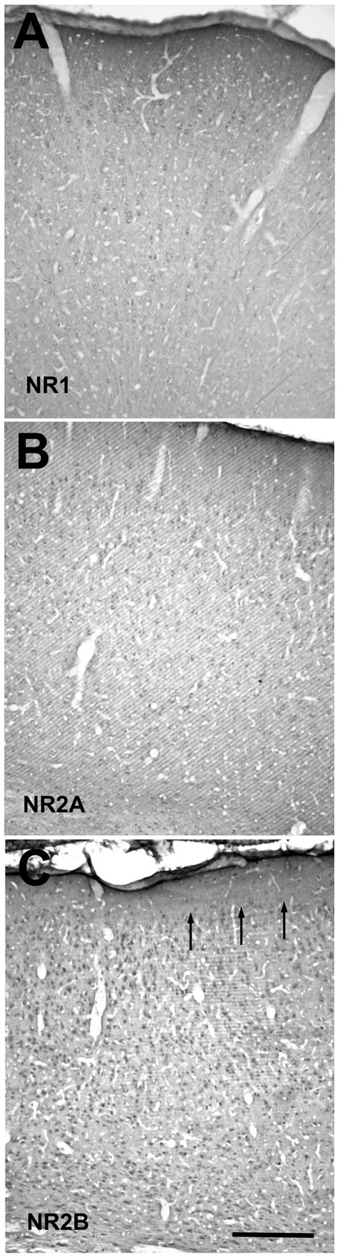
Light-microscopic view of coronal sections through the visual cortex of a P43 ferret, stained with NR1 (A), NR2A (B), or NR2B (C) antibodies. Staining is representative of that seen in all of the ages examined and does not reveal any laminar differences. Arrows in C mark the border between layers 1 and 2. Scale bar, 500 μm.