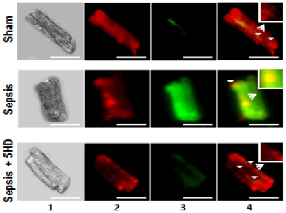
Figure 5. Effect of 5HD on mitochondrial membrane potential (ΔΨm) in ARVMs.
Mitochondrial ΔΨm was examined in sham and septic ARVMs and visualized using a fluorescent microscope. Representative photomicrographs (magnification 40×; scale bar, 75 µm) of sham and septic ARVMs treated in the presence and absence of 5HD are stained with JC-1 reagent. Lane 1 exhibits the ARVMs studied under a light microscope. The red (JC-1 aggregates in the mitochondria; lane 2) and green (JC-1 monomers in cytoplasm; lane 3) fluorescence was recorded and merged using an image software (lane 4). The white arrowheads depict the location of mitochondria in the ARVMs (which were zoomed 10 times and shown in the boxedsquare).

