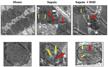
Figure 8. Ultrastructural changes in the mitochondrial membrane in LV tissue.
A. Ultrastructural changes in the mitochondrial cristae (using TEM) in the left ventricular tissue section (N = 5 in each treatment group) obtained from sham and septic rat hearts(6 and 12 hr post-sepsis). The mitochondrial cristae deformation was seen in the purified mitochondrial preparation from the septic rat left ventricular tissue (6 hr post-sepsis). B. The lower panel represents the magnified image of selected mitochondria in the purified mitochondrial preparation (red box, upper panel). Arrows points to the mitochondrial cristae deformation (RED) with balloon expansion (YELLOW). Magnification, 40 k×; Scale bar = 0.1 µm.

