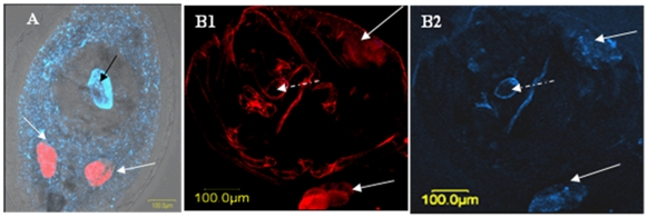
Figure 2. FISH of Bemisia tabaci nymphs parasitized by Eretmocerus mundus.
The procedure was performed using Portiera-specific probe (red) and Rickettsia-specific probe (blue). (A) Scattered (S) localization pattern. White arrows indicate bacteriocytes. Blue-speckled area is the whitefly hemocoel, and the dark, clear area corresponds to the outline of the roughly spherical Eretmocerus larva. Bright blue area (black arrow) shows the wasp larval gut. (B) Confined (C) localization pattern. (B1) Red area (white arrows) shows Portiera in the bacteriocytes (B2) Blue area (white arrows) shows Rickettsia in the bacteriocytes. The pictures of Rickettsia and Portiera are presented separately because of the faint signal seen by the former. Other blue and red areas in the pictures are due to autofluorescence of the whitefly nymph and the shell of the wasp's hatched egg (white dashed arrow).

