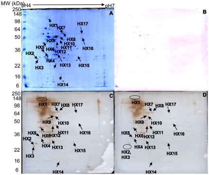
Figure 2. 2-DE gel and Western blot analyses of HA9801.
(A) HA9801 total cell proteins (pH 4–7, 13 cm), stained with colloidal Coomassie brilliant blue G-250. (B) 2-DE blot of S. suis stained with Ponceau S. (C) 2-DE blot of S. suis proteins probed with untreated antiserum. (D) 2-DE blot of S. suis proteins probed with “pre-absorbed” antiserum.

