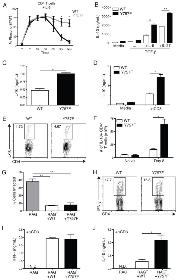
Figure 5.
Gp130 Y757F T cells produce augmented levels of IL-10 in response to IL-6 and IL-27. Purified CD4+ T cells from naïve WT and Y757F mice were stimulated in vitro with IL-6 (A) for various time points before being fixed, permeabilized and stained for intracellular phosphorylated STAT3. Data are pooled from 5 independent experiments. *p<.01. B) Purified CD4+ T cells from naïve WT or gp130 Y757F mice were activated with anti-CD3 and anti-CD28 for 48 hours in the presence of TGF-β with IL-6 or IL-27. IL-10 levels in culture supernatants were determined by ELISA. Data are representative of 4 independent experiments with similar results. **p<.001. C) IL-10 levels in the serum of infected WT (5) or gp130 Y757F (8) mice were determined by ELISA at day 8 post-infection. *p<.01. Data are pooled from two independent experiments. D) At day 8 post-infection, whole splenocytes from WT and Y757F mice were stimulated with media or anti-CD3 for 48 hours and assayed for IL-10 by ELISA. Data are representative of three independent experiments. *p<.01. D) Flow cytometry of IL-10 from splenocytes isolated from WT or gp130 Y757F mice at day 8 post-infection. Gated on CD3+ CD4+ events, cells were incubated with PMA, Ionomycin and BFA for 4 hours before being stained. F) The total number of IL-10+ CD4+ T cells was calculated in WT (n=5) and Y757F mice (n=6) at 8 days post-infection. Data are pooled from two independent experiments. *p<.01. G) 10 million splenic CD4+ and CD8+ T cells were isolated from WT and Y757F mice and transferred into RAG KO mice. The recipient RAG mice that received T cells from WT (n=5) or gp130 Y757F mice (n=5) were infected with T. gondii and at 8 days post-infection the percentage of infected PECs was enumerated. **p<.001. H) Flow cytometry of IFN-g from splenocytes isolated from RAG KO mice that received WT or gp130 Y757F mice at day 8 post-infection. Gated on CD3+ CD4+ events; cells were incubated with PMA, Ionomycin and BFA for 4 hours before being stained. I) At 8 days post-infection, whole splenocytes from recipient RAG KO mice were stimulated with anti-CD3 for 48 hours and assayed for IFN-γ by ELISA. J) At 8 days post-infection, whole splenocytes from recipient RAG KO mice were stimulated with anti-CD3 for 48 hours and assayed for IL-10 by ELISA. Data are representative of 3 independent experiments with similar results. *p<.01.

