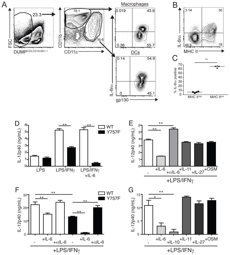
Figure 6.
IL-6 blocks the production of IL-12 by gp130 Y757F APC. A) Flow cytometry of IL-6 receptor alpha and gp130 expression on PECs from WT mice at day 4 post-infection. Gated on CD3/CD19/NK1.1neg, CD11c or CD11b+ cells. Representative FACS plot (B) and enumeration (C) of the percentage of MHC II negative or MHC II positive DCs that express the IL-6 receptor alpha. Data are representative of two independent experiments with similar results. D) Purified splenic DC from naïve WT and Y757F mice were stimulated in vitro with LPS, IFN-γ and IL-6 for 24 hours before IL-12p40 was measured in culture supernatants by ELISA. E) Purified splenic DC from naïve gp130 Y757F mice were stimulated in vitro with LPS, IFN-γ and IL-6, IL-11, IL-27, Oncostatin M or anti-IL-6 antibodies for 24 hours before IL-12p40 was measured in culture supernatants by ELISA. N=3 samples per condition. Data are representative of 3 independent experiments. F) Bone marrow-derived macrophages from WT and Y757F mice were stimulated in vitro with LPS, IFN-γ and IL-6 or anti-IL-6 antibodies for 24 hours before IL-12p40 was measured in culture supernatants by ELISA. G) Bone marrow-derived macrophages from gp130 Y757F mice were stimulated in vitro with LPS, IFN-γ and IL-6, IL-10, IL-11, IL-27 or Oncostatin M for 24 hours before IL-12p40 was measured in culture supernatants by ELISA.

