
Fig. 5. Recombinant viral infection of moDCs.
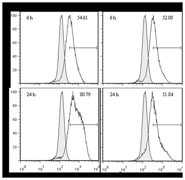
MoDCs were infected with hMPV, either WT or ΔG, and stained for specific cell surface antigens (anti-CD11c and MHC II), and for intracellular viral antigens. Top panels are results at 6 h p.i. and bottom panels represent 24 h p.i. Results are presented as histogram overlays of anti-hMPV staining versus isotype control. Results are representative of three separate experiments.