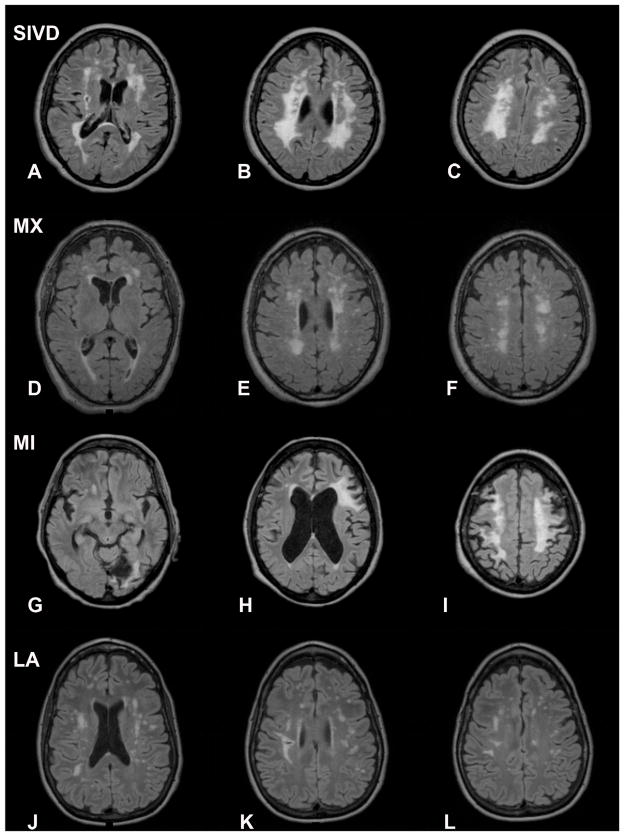
Figure 1.
FLAIR MRI scans from representative patients in the different subgroups. A–C) Patients in the subcortical ischemic vascular disease (SIVD) group show extensive white matter hyperintensities (WMHs) in a relatively symmetric distribution. D–F) Mixed VCI and AD (MX) patients have WMHs that are also symmetric. G–I) Multiple infarct (MI) patients have asymmetric lesions consistent with strokes. J–L) Leukoaraiosis (LA) patients have different patterns of WMHs that are difficult to characterize.

