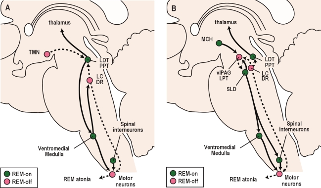
Figure 5.
Pathways that control REM sleep. (A) A classic perspective on REM sleep control involves interactions between the cholinergic and aminergic systems. REM sleep-active cholinergic neurons in the LDT/PPT activate thalamo-cortical signaling and drive atonia by exciting neurons in the ventromedial medulla that inhibit motor neurons. During REM sleep, monoaminergic neurons including the LC, DR, and TMN become silent, which disinhibits the LDT/PPT and lessens the excitation of motor neurons by NE and 5-HT. (B) Recent observations have expanded on the classic view of REM sleep control. In this model, mutual inhibition between REM sleep-on neurons of the sublaterodorsal nucleus (SLD) and REM sleep-off neurons of the ventrolateral periaqueductal gray and lateral pontine tegmentum (vlPAG/LPT) is thought to regulate transitions into and out of REM sleep. During REM sleep, SLD neurons activate GABA/glycine neurons in the ventromedial medulla and spinal cord that inhibit motor neurons. At most times, the vlPAG/LPT inhibits the SLD, but during REM sleep, the vlPAG/LPT may be inhibited by neurons making melanin concentrating hormone (MCH) and other neurotransmitters. Solid lines depict pathways active during REM sleep, while dashed lines are pathways inactive during REM sleep.

