Figure 5.
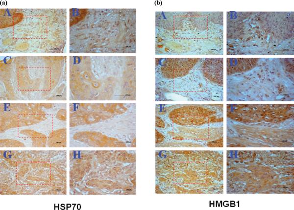
(a) Expression of HSP70 in ESCC tissues examined by IHC. Tissue-array slide was stained with polyclonal anti-HSP70 antibody at a 1:500 dilution. A: A representative normal esophagus tissue was negatively stained with anti-HSP70 antibody; C, E and G: Representative ESCC grade I, II and III tissues were positively stained with anti-HSP70 antibody (magnification,×100). B, D, F and H: The corresponding area (rectangle) of A, C, E and G was enlarged (magnification, ×200). (b) Expression of HMGB1 in ESCC tissues examined by IHC. Tissue-array slide was stained with polyclonal anti-HMGB1 antibody at a 1:500 dilution. A: A representative normal esophagus tissue was negatively stained with anti-HMGB1 antibody; C, E and G: Representative ESCC grade I, II and III tissues were positively stained with anti-HMGB1 antibody (magnification, ×100). B, D, F and H: The corresponding area (rectangle) of A, C, E and G was enlarged (magnification, ×200).

