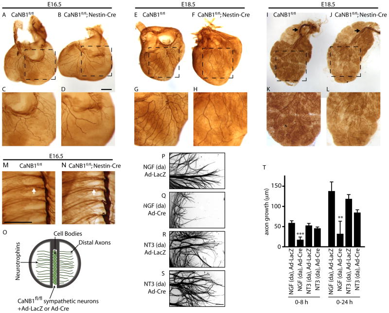
Figure 1. Calcineurin is required for NGF, but not NT-3-mediated axon growth in sympathetic neurons.
(A–L) Whole mount TH immunostaining shows reduced sympathetic fibers in target tissues in CaNB1fl/fl;Nestin-Cre mice as compared to CaNB1fl/fl controls, at E16.5 (heart: A–D), and E18.5 (heart: E–H and salivary glands: I–L). Higher magnification images are shown in the lower panels. Black arrows in I, J indicate sympathetic fibers approaching the salivary glands. Scale bar: 500 μm. (M–N) There are no differences in sympathetic chain organization between E16.5 CaNB1fl/fl (M) and CaNB1fl/fl;Nestin-Cre (N) mice. White arrow indicates TH-positive sympathetic fibers extending from sympathetic ganglia in both wild-type and mutant mice. Scale bar: 500 μm. (n=2 embryos for each genotype at E16.5, and at E18.5) (O) CaNB1fl/fl sympathetic neurons were infected with adenoviral vectors expressing Cre (Ad-Cre) or LacZ (Ad-LacZ). Neurotrophins were added only to distal axons (da). (P–S) Cre-mediated calcineurin deletion specifically decreases NGF, but not NT-3-mediated axon growth. Axons were stained with β-III-tubulin for visualization after quantification of axon growth. Scale bar, 80μm. (T) Quantification of axon growth in compartmentalized cultures over 0–8 hr and 0–24 hr, ** p<0.01, ***p<0.001. Results are mean ± SEM from n=5 experiments.

