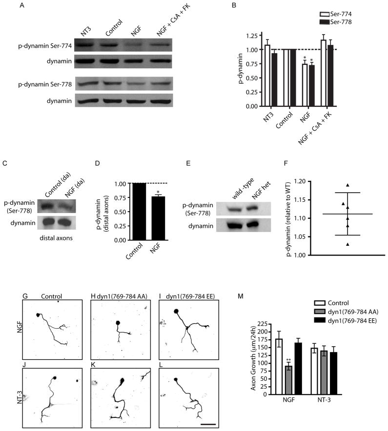
Figure 5. NGF promotes axon growth through dynamin dephosphorylation.
(A) NGF stimulation results in dephosphorylation of dynamin1 in a calcineurin-dependent manner. Neuronal lysates were immunoblotted using phospho-Ser774 and phospho-Ser778 dynamin antibodies. Immunoblots were stripped and reprobed for total dynamin1. (B) Densitometric quantification of phospho-dynamin1 levels *p<0.05, n=6. (C) NGF stimulation results in dephosphorylation of dynamin1 (Ser 778) in distal axons. Immunoblots were reprobed for total dynamin1. (D) Densitometric quantification of phospho-dynamin1 (Ser778) in axons, *p<0.05, n=3. (E–F) NGF+/− mice have increased levels of phospho-dynamin1 in sympathetic axons in vivo. Salivary gland lysates from P0.5 wildtype and NGF+/− mice were immunoblotted using phospho-dynamin1 (Ser778) antibody. Immunoblots were reprobed for total dynamin1. (F) Densitometric quantification of phospho-dynamin1 (Ser778) after treatments as described in (E), represented as a scatter plot with 95% confidence intervals. n=6 pups for each genotype. (G) Dephosphorylation-dependent dynamin1 function is required for NGF-mediated axon growth. Introduction of dyn1(769-784 AA) (H) but not the dyn1(769-784 EE) (I) peptide decreased NGF-dependent axon growth over 24 hr. NT-3-mediated growth was unaffected by introduction of dyn1 phosphopeptides (J–L). Scale bar, 100μm. (M) Quantification of axon growth. **p<0.01, n=3.

