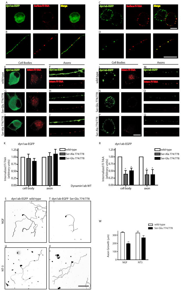
Figure 7. Phosphoregulation of dynamin1ab is required for TrkA endocytosis and axon growth.
(A–D) Dynamin1aa isoform (A,B) shows diffuse cytoplasmic localization while dynamin1ab isoform (C,D) shows punctate localization in cell bodies and axons. FLAG immunostaining shows surface FLAG-TrkA receptors in cell bodies (A,C) and in axons (B,D). Scale bar, 10 μm. (E–J) Phosphoregulation of dynamin1aa is not required for NGF-dependent TrkA internalization. Neurons were transfected with FLAG-TrkA and either wild-type dynamin1aa-EGFP (E, F), Ser-Glu 774/778 dynamin1aa-EGFP (G,H), or Ser-Ala 774/778 dynamin1aa-EGFP (I,J). Cell bodies are shown in E, G, I. Axons are shown in F, H, J. Scale bar, 10μm. (K) Quantification of NGF-dependent TrkA internalization in cell bodies and axons. n=3. (L–Q) Phosphomutants of dynamin1ab disrupt NGF-dependent internalization of TrkA. Neurons were transfected with FLAG-TrkA and either wild-type dynamin1ab-EGFP (L,M), Ser-Glu 774/778 dynamin1ab-EGFP (N,O), or Ser-Ala 774/778 dynamin1ab-EGFP (P,Q). Cell bodies are shown in L, N and P. Axons are shown in M, O and Q. Scale bar, 10μm. (R) Quantification of internalized TrkA. *p<0.01, n=3. (S–V) Phosphoregulation of dynamin1ab is required for NGF-, but not NT-3-, dependent axon growth. NGF-mediated growth is blocked in sympathetic neurons expressing dynamin1ab-EGFP Ser-Glu 774/778 (T) as compared to wild-type dynamin1ab-EGFP (S). NT-3-mediated growth was unaffected (U,V). (W) Quantification of neurite length. **p<0.01, n=3. Scale bar: 50 μm.

