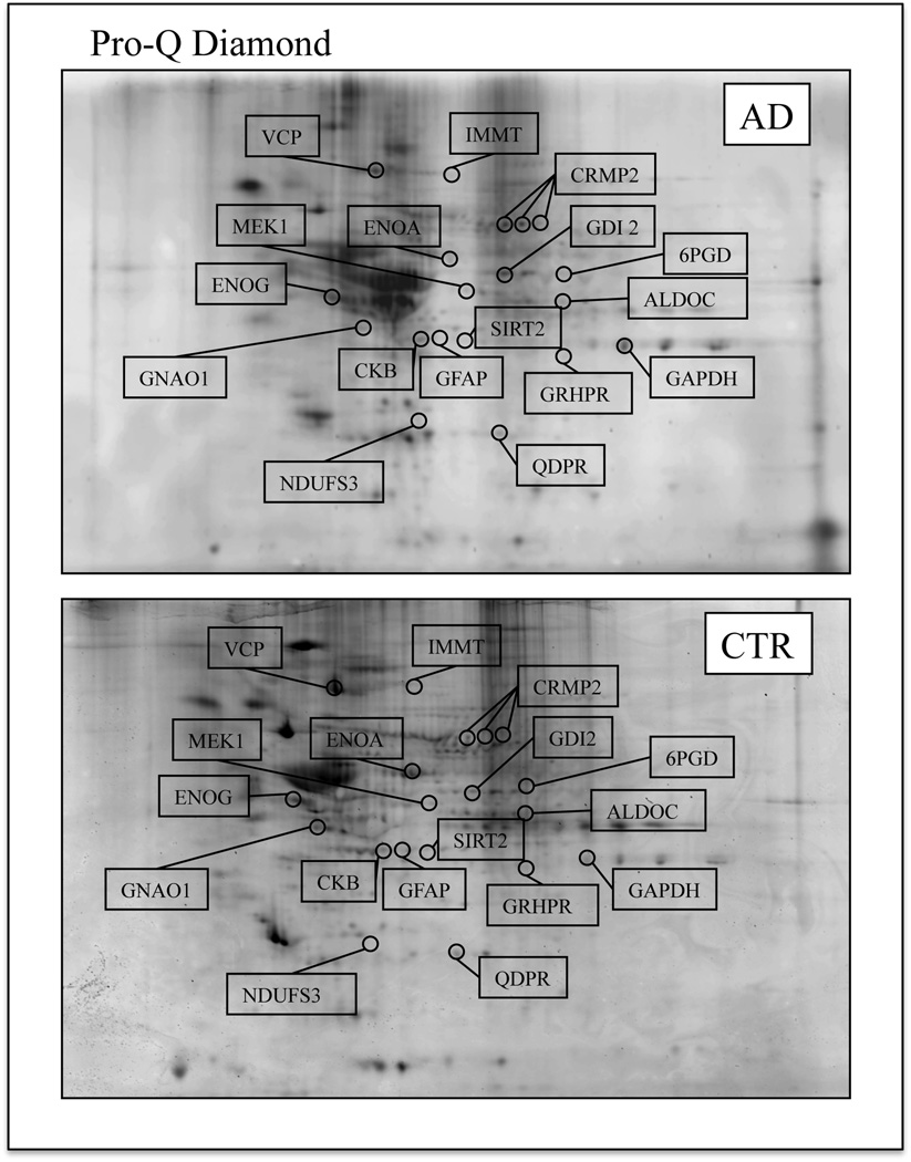
Figure 1.
Representative 2D phosphorylation maps of AD (top) and CTR (bottom) hippocampus. Gels were stained using Pro-Q Diamond fluorescent dye. The spots showing altered phosphorylation levels between AD and control are labeled. Relative change in phosphorylation, after normalization of the immunostaining intensities to the protein content, was significant for 17 spots.

