
FIGURE 1. In vivo metabolite analysis of 18F-FAC.
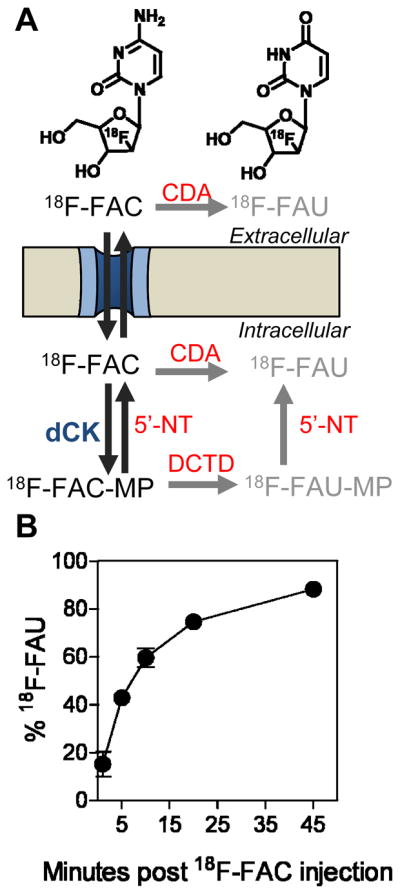
(A) Chemical structures of 18F-FAC and 18F-FAU and schematic showing the extracellular and intracellular routes by which 18F-FAC can be deaminated to 18F-FAU. CDA, cytidine deaminase; 5′-NT, 5′nucleotidase; DCTD, cytidylate deaminase (catabolic enzymes are shown in red fonts); (B) Percentage of total detected radioactivity in plasma that is attributable to 18F-FAC over time as determined by HLPC analysis. Analysis is carried out following intravenous injection of 18F-FAC in mice.