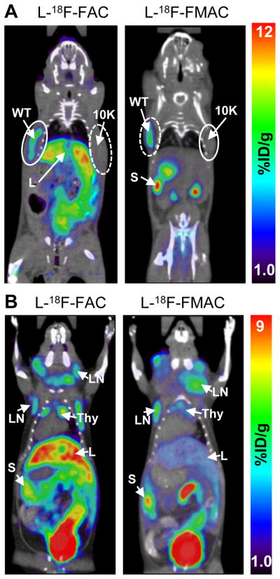
FIGURE 6. L-18F-FAC and L-18F-FMAC microPET/CT scans of malignant and autoimmune lymphoproliferative disorders.
(A) L-18F-FAC and L-18F-FMAC microPET/CT imaging of L1210 lymphoma tumors. The L1210 parental cell line (WT, solid-lined circle) and the dCK-deficient variant L1210-10K (10K, dash-lined circle) were injected subcutaneously under the left and right shoulder of the mouse, respectively. Only the parental cell line accumulated both probes; (B) L-18F-FAC and L-18F-FMAC microPET/CT imaging of autoimmune B6.MRL-Faslpr/J mice. Both probes detected cervical, axillary and brachial lymphadenopathy in these mice. L, liver; LN, lymph nodes; S, spleen; Thy, thymus; number of mice per probe ≥ 3.

