Figure 1.

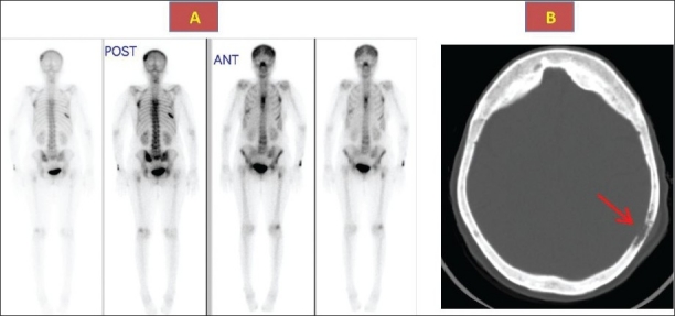
Bone scan (1a) showing foci of increased activity in the ribs, skull, spine and CT skull (1b) showing thickened sclerotic and lytic lesions (arrow) consistent with Paget disease.

Bone scan (1a) showing foci of increased activity in the ribs, skull, spine and CT skull (1b) showing thickened sclerotic and lytic lesions (arrow) consistent with Paget disease.