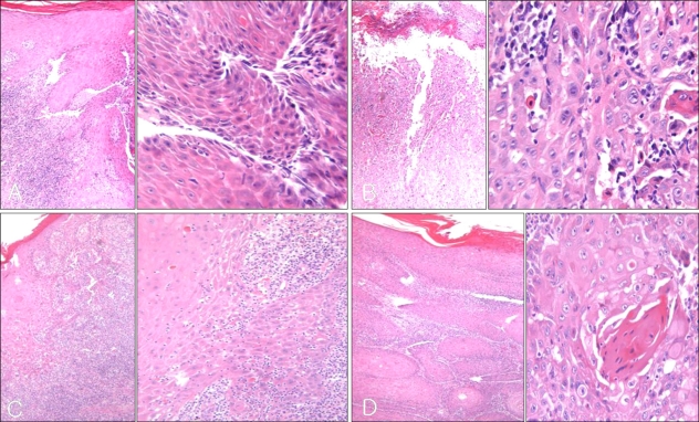
Fig. 2.
Histology of our patient. (A) Chronic inflammatory cell infiltration in the dermis without epidermal change at the initial visit. (B) Atypical, pleomorphic keratinocytes are confined to the epidermis 30 months later. (C) Extension of atypical keratinocytes in the tumor nest beyond the basement membrane after excision. (D) Suggestive squamous cell carcinoma in the lymph node (A, B, C, D: H&E, ×40, ×400).

