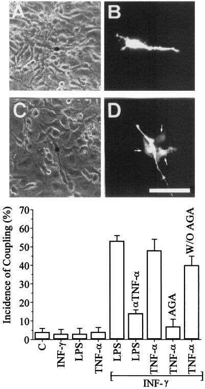
Figure 2.
Cytokines induce dye coupling between cultured rat microglia. (A and C) Phase-contrast views of the fluorescence fields shown in B and D, respectively. (B) An example of lack of dye coupling observed under control conditions. (D) A cell that is dye coupled to three neighboring cells in a microglia culture treated with 1 ng/ml INF-γ plus 1 ng/ml TNF-α for 9 h. (Bar, 80 μm.) (Graph) The incidence of dye coupling (Lucifer yellow) was evaluated in cultures of rat microglia under control conditions and after cytokine treatment for 9 h. Treatment with 1 ng/ml INF-γ, 1 μg/ml LPS, or 1 ng/ml TNF-α did not increase coupling above control levels. INF-γ (1 ng/ml) plus LPS (1 μg/ml) caused a large increase in coupling, which was largely prevented by cotreatment with an anti-TNF-α antibody. Treatment with 1 ng/ml INF-γ plus 1 ng/ml TNF-α also caused a large increase in coupling; this coupling was blocked by 35 μM 18α-glycyrrhetinic acid (AGA), and the block was reversed after the blocker was washed out (W/O AGA). Each histogram bar corresponds to the mean ± SD of seven experiments, in each of which a minimum of 10 cells were scored.

