
Figure 7. Conformational states of methionine synthase.
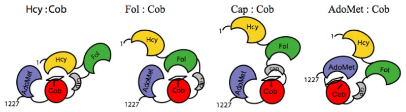
The four modules are shown in gold (Hcy-binding), green (folate-binding), red (cobalamin-binding domain) and gray (cap domain), and blue (AdoMet-binding). The corrin ring of methylcobalamin is indicated by the rectangle on top of the cobalamin-binding domain, and His759 is indicated by the vertical line. In the AdoMet:Cob conformation, the histidine is displaced as indicated in the cartoon, and the corrin ring tilts away from the cobalamin-binding domain and from Cα of His 759. Reprinted from 20.