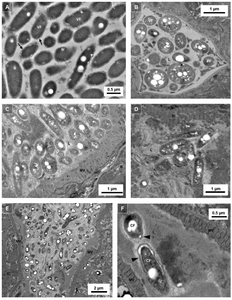
Figure 4.

Intraluminal Legionella exhibit morphologically differentiated forms. Transmission electron microscopic images of (A) Legionella harvested from 6-day old assay plate culture without the presence of N2 nematodes; (B) cross-cut pharynx section of N2 nematodes colonized with L. longbeachae ATCC 33462 (note triangular shape of the grinder); (C) and (D) mid-body cross-cut digestive tract sections of N2 nematodes colonized with L. pneumophila Philadelphia-1 type strain; and (E) and (F) mid-body transverse-cut digestive tract sections of nematodes colonized with L. pneumophila SVir. Note that the thickened cell walls and the white circular spaces within the Legionella bacteria are poly-β-hydroxybutyrate (PHBA) granules. VF, vegetative form; CF, cyst-like form; L, intestinal lumen; IC, intestinal cell; MV, microvilli; arrow, thin cell wall of VF Legionella; arrowhead, thick cell wall of CF Legionella.
