Figure 5.

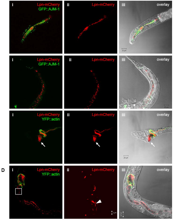
Presence of Legionella in nematodes cultivated in a soil environment. Confocal microscopic images of mCherry-producing L. pneumophila KB290 in C. elegans PS3729 (A) head of a L2 stage and (B) tail of a young adult 30 days after initial inoculation, and in WS1904 (C) tail of a L3 stage and (D) mid-body of an adult views 60 days after initial inoculation. Panels i-iii correspond (i) red and green channels showing fluorescent KB290 and nematode intestinal cells, (ii) red channel alone showing only KB290,and (iii) overlayed red channel, green channel, and DIC bright-field images. Green fluorescence indicates the apical tight junctions within the pharynx structure in PS3729 and outlines the intestinal tract in WS1904. Note that image (Dii) is at higher magnification and corresponds to the outlined inset box in (Di). Arrows denote a heavily colonized anus leading to anal swelling; arrowhead demonstrates bacilli that appear to have recently divided.
