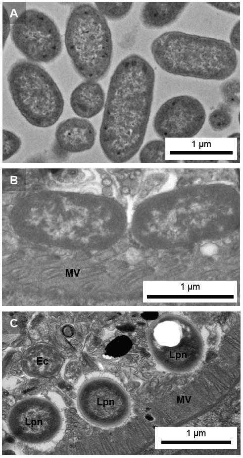
Figure 6.

Ultrastructural analysis of bacteria within C. elegans digestive tract. Transmission electron microscopic images of (A) plate grown E. coli OP50, and transverse-cut sections of the digestive tracts of C. elegans PS3729 nematodes extracted from a soil environment 90 days after initial inoculation with intermittent supplementation of E. coli OP50 detail bacteria embedded within intestinal lumen lining the digestive tract; (B) E. coli OP50, and (C) mixed population of OP50 and Legionella. Note ultrastructural differences between OP50 and Legionella, in particular the thickened cell walls and the white spaces indicating the presence of PHBA in Legionella. Lpn, L. pneumophila; Ec, E. coli OP50; MV, microvilli.
