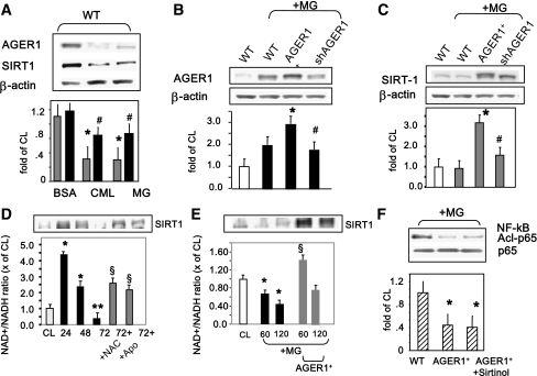
Figure 2.
AGEs suppress AGER1, SIRT1 protein, and NAD+ levels as well as NF-κB p65 deacetylation in THP-1 cells. A: Western blots (upper panels) and densitometry (lower panels) results are shown for AGER1 (black bars) and SIRT1 (gray bars) protein expression in THP-1 cells (wild-type [WT]) stimulated with CML-BSA (150 μg/mL), MG-BSA (60 μg/mL), and BSA (60 μg/mL) for 72 h. B and C: MG-induced effects on SIRT1 are AGER1-dependent. WT or THP-1 cells transfected with AGER1 (AGER1+) or short-hairpin RNA for AGER1 (shAGER1) were stimulated by MG (60 μg/mL) for 24 h before Western blots (upper panels) and densitometry plots (lower panels; AGER1, black bars; SIRT1, gray bars; WT, open bars). Data (mean ± SEM) of three to five experiments, derived from test/β-actin ratio, are shown as fold above control (cells alone, WT). *P < 0.001 vs. BSA or cells alone. #P < 0.002 vs. maximal values. D and E: AGE-induced effects on SIRT1 are NAD+-dependent and regulated by OS (D) and AGER1 (E). THP-1 cells were cultured with MG-BSA (60 μg/mL) or media (CL) for up to 72 h prior to Western blotting for SIRT1 (top inset) and NAD+/NADH ratio in the presence or absence of antioxidants (NAC or apocynin) in WT or AGER1+-transduced cells (E). NAD+/NADH ratio is shown as fold (mean ± SE) above control (n = 3, each in triplicate). *P < 0.001 vs. control. §P < 0.002 vs. maximal increase. F: NF-κB p65 hyperacetylation is induced by AGEs but is blocked by AGER1. Acetyl-p65 was determined in THP-1 cells, after MG stimulation (60 μg/mL) for 72 h in the presence or absence of SIRT1 inhibitor, sirtinol (10 μmol/L). Western blots and density plots are shown as mean ± SEM from four independent experiments. *P < 0.002 vs. nonstimulated or vs. nontransduced THP-1 cells.

