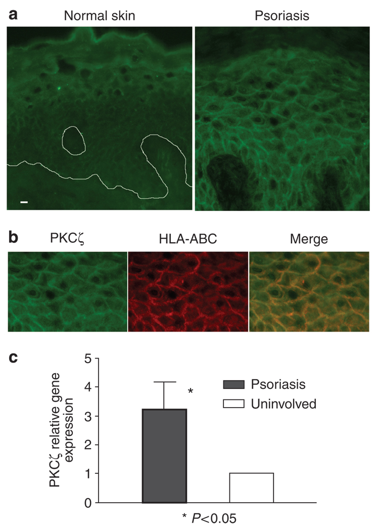
Figure 4. Increased membrane expression of PKCζ by psoriatic plaques.
(a) Immunofluorescence of PKCζ in psoriasis and normal control skin showed increased cytoplasmic and membrane staining for PKCζ in psoriasis. Bar = 10 µm. (b) Double labeling with anti-HLA-ABC antibody showed membrane localization of PKCζ in psoriasis lesion. (c) When mRNA from six pairs of both lesional and uninvolved skin specimens was examined by real-time PCR, PKCζ gene expression was increased in all psoriasis samples compared with uninvolved skin (P < 0.05, Wilcoxon rank test). Values are mean ± SD, error bar = 1 SD.

