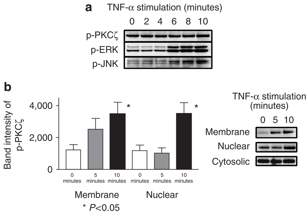
Figure 7. Increased phosphorylated PKCζ in membrane fractions of KCs stimulated with TNF.
(a) KCs were stimulated with 100 ng ml−1 of TNF-α for 0, 2, 4, 6, 8, and 10 minutes. While no significant change in the quantity of phospho-PKCζ was observed, pERK and pJNK were increased 6 minutes after TNF-α stimulation. (b) Increased phospho-PKCζ in the membrane and nuclear, but not in the cytosolic fractions, of primary KCs stimulated with TNF-α 100 ng ml−1 for 5 and 10 minutes (Student’s t-test). These blots are representations of three separate experiments. Values are mean ± SD, error bar = 1 SD.

