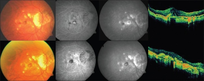
Figure 5.

Top (left to right): Pretreatment color fundus photograph, fluorescein angiography (early and late phases) showing active leakage and optical coherence tomography revealing increased retinal thickness in a case in group 5 (reduced-fluence photodynamic therapy with ranibizumab). Bottom (left to right): Post-treatment color fundus photograph, fluorescein angiography (early and late phases) showing resolution of the leakage with staining of the scar and optical coherence tomography showing a hyper-reflective scar with resolution of fluid in the same case mentioned above
