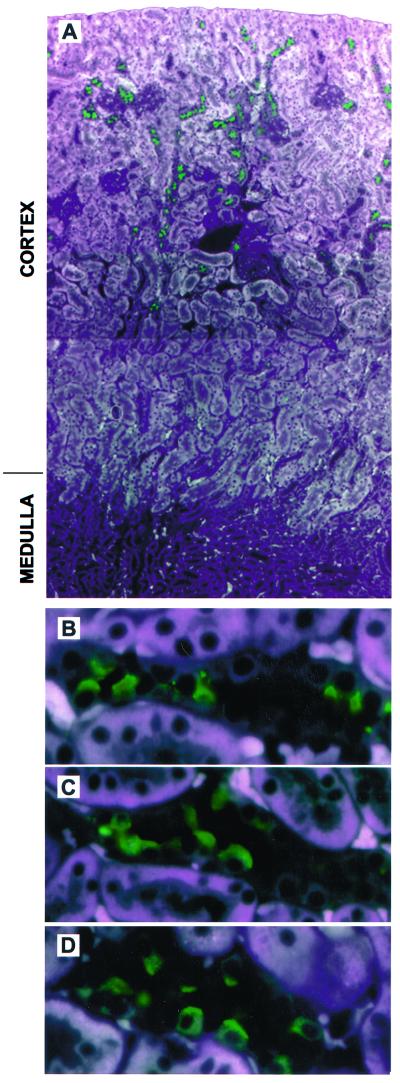
Figure 2.
Immunofluorescent staining of pendrin in mouse and rat kidney. Paraformaldehyde-fixed, paraffin-embedded kidney sections were incubated with rabbit anti-pendrin antibodies followed by fluorescein-labeled anti- rabbit secondary antibody (1:250). (A) A low-power composite view (original magnification ×100) of mouse kidney showing cortical cells staining with an anti-pendrin antibody (h766–780; 1:1000). Strong pendrin-specific staining (in green) is seen in the cortex; no staining is detected in the outer or inner medulla. (B) A higher-power view (original magnification ×630) of the section shown in A illustrating the apical staining of pendrin-positive cells. Note that pendrin is only detected in a subset of cells within a given tubule. Similar studies were performed with rat kidney stained with the same antibody as A and B (C) or with a published (7) anti-pendrin antibody (D; r612–625; 1:1000), revealing an identical pattern of apical staining in the cortex. All photomicrographs represent merged images captured with three distinct excitation lights (345, 490, and 540 nm); this approach allows the cellular structure to be visualized in conjunction with the FITC-associated staining (in green). Note that the white areas in A reflect fluorescence of the proximal tubules; the same background is also seen in the presence of immunizing peptide as well as in kidney sections derived from Pds-knockout mice.

