Figure 1.
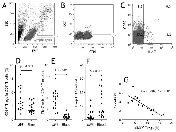
Both CD39+ regulatory T (Tregs) cells and Th17 cells increased in malignant pleural effusion (MPE). (A) Lymphocytes were identified based on their characteristic properties shown in the forward scatter (FSC) and sideward scatter (SSC). (B) A representative gating was set for CD4+ T cells from pleural lymphocytes. (C) A representative dot plots showing expression of CD39 and IL-17 in pleural CD4+ T cells. Comparisons of percentages of CD39+Tregs (D), Th17 cells (E), and ratios of CD39+Tregs/Th17 cells (F) in MPE and blood from patients with lung cancer (n = 16). The percentages of CD39+Tregs and Th17 cells were determined by flow cytometry. Horizontal bars indicate medians. Comparison was made using a Wilcoxon signed-rank test. (G) The percentages of Th17 cells correlated with CD39+Tregs cells in MPE. Correlations were determined by Spearman's rank correlation coefficients.

