
Figure 10.
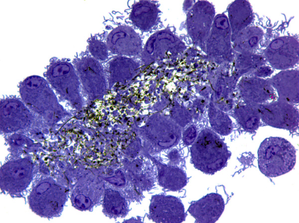
Morphology of granuloma illustrating secondary agglomeration of carbon nanotubes by bone marrow-derived macrophages exposed initially to well-dispersed materials. After 14 days, cells were fixed in situ, embedded in plastic, and sectioned at 0.5 μm, and stained with toluidine blue for light microscopy. Magnification: 1000×.