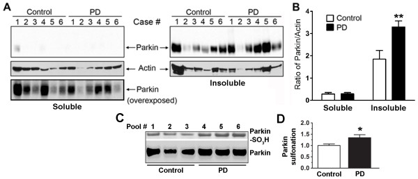
Figure 6.
Selective increase in insoluble parkin levels and sulfonation in idiopathic PD brains. (A) The "Soluble" and "Insoluble" fractions of brain tissue lysates from the caudate nucleus were blotted for parkin immunoreactivity with PRK8 monoclonal antibody. Immunoblotting revealed that the levels of parkin were significantly increased in the caudate of sporadic PD patients compared to controls. (B) Relative amount of parkin normalized to actin. Data are expressed as mean ± SEM, n = 6; **p < 0.01 by post-hoc ANOVA. (C) The "Insoluble" fraction of tissue lysates from postmortem human brains of either normal control subjects or sporadic PD cases were immunoblotted for parkin sulfonation. Pooled brain tissue lysates were obtained from 12 patient samples (see subject information in Additional file 1, Table S2). Increased parkin sulfonation was observed in PD compared to Control. (D) Quantification of parkin sulfonation by normalizing the intensity of sulfonated parkin to total parkin. Data are expressed as mean ± SEM, n = 3; *p < 0.05 by post-hoc ANOVA.

