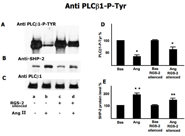
Figure 4.
Effect of Ang II on PLCβ1-Tyr-phosphorylation and SHP-2 association in RGS-2 silenced and not silenced cells. RGS-2 not silenced (lanes a, b) and silenced (lanes c, d) fibroblasts were incubated with Ang II (lanes b, d,) or with vehicle (lane a, c) for 1 hour as described in the Methods. Immunocomplexes were isolated and analyzed as described in methods. Panel A is PLCβ1 Phospho-Tyrosine levels, panel B is SHP-2 protein levels and panel c is PLCβ1 protein levels. The figure is representative of five separate experiments carried out in duplicate. Panels D and E present the percent change relative to unstimulated cells and SD (N = 5 experiments) for panel A and B respectively. Panel D: •: p < 0.0001 vs Basal; *: p < 0.0001 vs Ang RGS-2 Silenced; **: p < 0.0001 vs Basal RGS-2 Silenced. Panel E: •: p < 0.0001 vs Basal; *: p = 0.04 vs Ang RGS-2 Silenced; **: p = 0.001 vs Bas RGS-2 Silenced.

