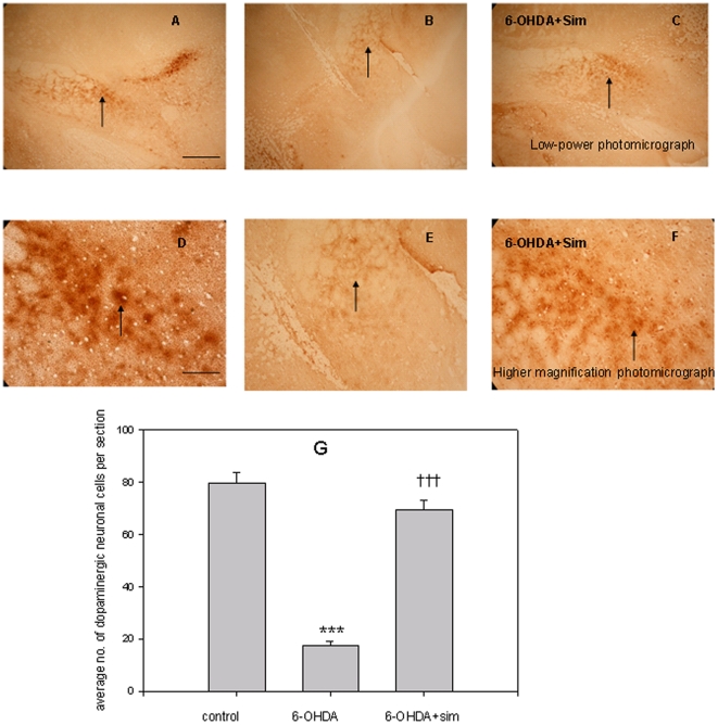
Figure 1. Effects of 6-OHDA lesion and simvastatin on TH immunohistochemistry staining in the SNpc.
Figs. A, B, C shows TH staining in low-power photomicrograph in the SNpc of unlesioned, 6-OHDA-lesioned, and 6-OHDA-lesioned with simvastatin treatment groups, respectively. Bar = 450 µm. Figs. D, E, F shows TH staining at higher magnification photomicrograph in the SNpc of unlesioned, 6-OHDA-lesioned, and 6-OHDA-lesioned with simvastatin treated groups, respectively. Bar = 120 µm. Fig. 1G represents the average number of TH-positive dopaminergic neurons in the SNpc of unlesioned (control), 6-OHDA lesioned, and 6-OHDA lesioned with simvastatin treatment groups. The values represent mean ±SEM, n = 6–8. ***p<0.001, 6-OHDA group versus control group; ††† p<0.001, 6-OHDA+simvastatin group versus 6-OHDA group.

