Figure 1.
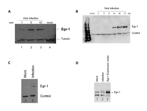
Egr-1 protein was induced by HSV-1 infection 24 hours post treatment. a.) This figure is a western blot analysis showing the expression of Egr-1 protein (83 Kda) in infected VERO cells 24 hours post treatment. MOI of 1, 5, and 10 were used and more viruses led to more Egr-1 production. Noted that infection at higher moi (Lane 3) led to cell death therefore no significant increase of Egr-1 production was observed. b.) Egr-1 was induced by HSV-1 infection (MOI-5) in VERO cells and detectable levels were observed at 24, 48, and 72 hours post treatment. c.) Egr-1 induction was also seen in SIRC cells infected with a MOI of 5. d.) Uninfected HEK 293 cells express Egr1 (lane 1), and no induction was seen upon infection with HSV-1 MOI 10 (lane 2). Overexpression of Egr-1 was seen in HEK293 cells transfected with Egr1 expression vector (lane 3).

