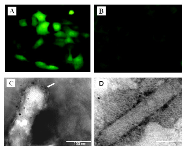
Figure 2.
Characterization of BV-Dual-HA. PK-15 cells were transduced with BV-Dual-HA (A) or AcMNPV-WT (B) at an MOI of 10. At 48 h post-transduction, cells were fixed with absolute methanol, and processed for indirect immunofluorescent assay. Bound antibodies were detected by FITC-labeled anti-mouse IgG by fluorescence microscopy (green). Original magnification × 200. Electron micrograph of recombinant baculovirus displaying HA on the viral envelope. BV-Dual-HA (C) and AcMNPV-WT (D) were treated with anti-HA monoclonal antibodies, followed by labeling with anti-mouse IgG-gold conjugate. One end of the viral envelopes was strongly labeled with gold particles (arrows). Bars - 100 nm.

