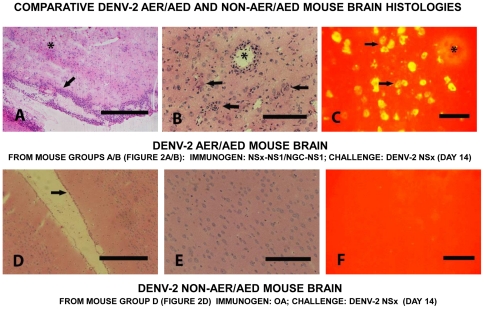
Figure 4. Comparative DENV-2 AER/AED and non-AER/AED mouse brain histologies.
Representative DENV-2 AER/AED mouse brain sections (A, B and C) from group A/B mice (Figure 2A/B) and DENV-2 non-AER/AED mouse brain sections (D, E and F) from group D mice (Figure 2D), immunized with either the NS1 glycoprotein of the DENV-2 NSx strain or ovalbumin by the combined i-p./s-c. routes, respectively, prior to challenged with the DENV-2 NSx strain by the i-c. route, collected on day 14 after challenge. Cryostat-cut sections were either stained with standard hematoxylin and eosin (H&E) (A, B, D and E) or tested in immuno-fluorescence antibody (IFA) assays using human PAbs reactive against the DENV E glycoprotein and FITC-labelled secondary PAbs. The DENV-2 AER/AED mouse meninges showed extensive mononuclear cell infiltration (arrowed) with microglial nodule formation and many eosinophilic-staining dead neurones (asterixed) (100x: 100 µm bar) in their brain parenchyma (A). The DENV-2 AER/AED mouse microglial nodules were located throughout their brain parenchyma (arrowed) and blood vessels showed perivascular cuffing with mononuclear cells (asterixed) (200x: 50 µm bar) (B). DENV-2 E glycoproteins were present in the microglial cells, including those which formed nodules (arrowed) throughout their brain parenchyma, but not in the mononuclear cells in the perivascular infiltrate (asterixed) (400x: 20 µm bar) (C). In contrast, the DENV-2 non-AER/AED mice showed normal meninges (arrowed) (100x: 100 µm bar) (D) and no pathological changes in their brain parenchyma (200x: 50 µm bar) (E) and no DENV-2 E glycoproteins were present in these cells (400x: 20 µm bar) (F).

